

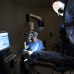
Start of Surgery
We are done preparing. Time to get to work:
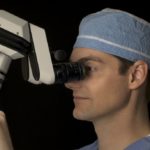
The microscope was moved back into position…
Cataract surgery is microsurgery. Without a microscope it would not be possible to complete the steps to follow.
…and a paracentesis was created at the one and five o’clock positions…
A ‘paracentesis’ is a small incision (usually 1.0mm wide) in the cornea that allows the surgeon to place instruments or inject fluids into the eye (more on that next). In general when discussing orientation durging surgery the eye is compared to a clockface with 12:00 the uppermost portion of the cornea (near the upper eyelid or brow) and 6:00 being the lowermost portion (near the lower eyelid or feet).
…through which 0.14 cc of Epi-Shugarcaine was injected into the anterior chamber.
‘Epi-Shugarcaine’ is a sterile solution of anesthetic and dilating medications developed by the late Dr. Joel Shugar. Not all surgeons use this solution. However, it can result in better anesthsia and dilation. I do not use it in all cataract surgeries but if a patient is on Flomax or has a small pupil I will instill Epi-Shugarcaine.
The ‘anterior chamber’ is a clinical term for the space between the iris (the colored part of the eye) and the posterior (backside of the) cornea (the clear front part of the eye on which a contact lens sits).
Viscoat was then injected into the anterior chamber firming up the globe.
Viscoat and Provisc are just two of many brands of viscoelastic. A ‘viscoelastic’ material, aka ‘viscosurgical device’ is a gel-like material that is placed in the eye in order to (1) create and maintain space to work-in, (2) protect the corneal endothelium. he corneal endothelium is made up of cells that pump fluid out of the cornea (keeping it clear). When these cells absorb the phacoemulsification energy (decribed in a later post) it ‘shocks’ them resulting in ‘corneal edema’ or a thickening of the cornea. Although usually self-limited, if this edema does not go away the vision would be blurred and a corneal transplant might be necessary. Thus, you can see why we would want to use something to protect the corneal endothelium.
A clear cornea temporal incision was then created with a metal keratome…
In order to remove the cataract and later place a new lens in the eye an incision must be made in the cornea. Currently there is no way around this. Thus, cataract surgery requires an incision. That being said, the incision is usually very small – on the range of 2.2-3.5mm wide.
This incision can either be made with a very sharp metal or diamond blade. Either one would make a standard razor blade appear dull by comparison. Because these blades must be manufactured to very exacting specifications they are quite expensive. A disposible metal blade runs anywhere from $35-70 per knife. Diamond blades, on the other hand, can be used hundreds of times before needing to be repaired or replaced. However, they are exceedingly expensive ($1,100-4,000) and are easily dulled or damaged.
…following which a continuous curvilinear capsulorrhexis was created using a bent needle cystatome on a Provisc syringe followed by capsulorrhexis forceps.
This is considered by many surgeons to be the most challenging element of the surgery. In order to get to the cataract an opening must first be made in the ‘capsule’ a delicate film-like material that holds the lens in place. This material is very thin (measured in microns, or millionths of a meter) and transparent. It is held in place by cables called ‘zonules’ that stretch it out over the surface of the lens.
Ideally, the surgeon wants to make a circular, or ‘curvilinear’ opening in the capsule. However, as you can imagine, tearing an opening in a thin, clear material on stretch is not a task for the faint of heart (considering that if the tear extends beyond the edge of the lens the rest of the cataract surgery becomes challenging, at best). There are two main ways of doing this: with a bent needle cystatome or with forceps. I use both. My father is a mechanic and taught me to use the best tool for the task at hand. As such, I find that the cystatome works best to start the capsulorrhexis and the forceps give me the most control over the shape of the opening.
© 2009 David Richardson, MD


Leave a Reply